Anatomy Of Chest Bone : Rotation of 3D skeleton.ribs,chest,anatomy,human,medical ... : Compare the nuclear medicine scans to anatomical diagrams.. The two bones are joined at a slight angle that protrudes anteriorly (sternal angle, angle of louis). It describes the theatre of events. Learn about this topic at kenhub! Where is the sternum found. It is made up of the wrist joint, the carpal bones, the metacarpal bones, and the phalanges.
In this article we will focus on: The wrist consists of multiple joints where the bones of the arm and hand meet. The two bones are joined at a slight angle that protrudes anteriorly (sternal angle, angle of louis). Bone comprises the structure of the skeletal system and provides lever arms for locomotion. Diaphyseal bone is organized to create the best balance between weight and structural strength.
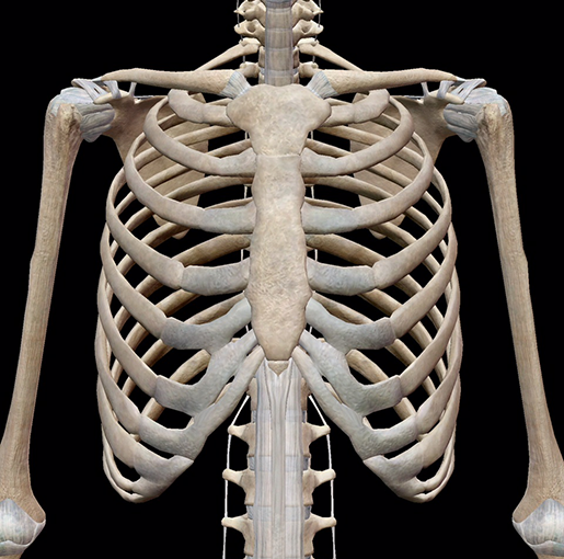
Bones of the Chest and Upper Back from www.innerbody.com Breast bone anatomy human breast bone anatomy bone anatomy chest. It is comprised of many bones, formed by intramembranous ossification, which are joined together by sutures (fibrous joints). Where is the sternum found. Language and terminology for the study of the anatomy of the thorax. These bones form by the thickening of a. Upper segment of sternum, flattened roughly triangular bone, o… the bony structure that forms the middle portion of the sternu… The interpretation of a chest film requires the understanding of basic principles. The former is a type of connective tissue made up of cells suspended in a matrix:
Your rib cage, for example, acts like a shield around your chest to protect important organs inside such as your lungs and heart.
These bones form by the thickening of a. These joints fuse together in adulthood. They are always longer than they are wide the vertebrae are irregular bones. Human anatomy detail of chest and shoulder. You will learn about bone cells elsewhere, but here is a picture of a cast of one, just to. Human chest bone structure parts of the chest bones. Irregular bones have complex shapes. Bone of chest and their parts. 400 x 300 jpeg 50 кб. This webpage presents the anatomical structures found on wrist mri. The name for the bone was derived from a deity of greek mythology called atlas, who supported the. 1300 x 1065 jpeg 178 кб. What can you label/identify on the nmt exam.
It supports the weight of the skull. Learn about each muscle, their locations & functional anatomy. The former is a type of connective tissue made up of cells suspended in a matrix: It can help you understand our world more detailed and specific. Compare the nuclear medicine scans to anatomical diagrams.
3D Skeletal System: Bones of the Thoracic Cage from www.visiblebody.com Human chest bone structure parts of the chest bones. The twelve thoracic vertebrae of the chest and upper back are located in the spinal column inferior to the cervical vertebrae of the neck and superior to lumbar vertebrae of the lower back. Spot views were taken of the chest, spine, hand, and foot. Identify the following structures on the lateral chest radiograph: This anatomical midline can be useful in assessing for symmetry in breast augmentation or in performing a median sternotomy. Upper segment of sternum, flattened roughly triangular bone, o… the bony structure that forms the middle portion of the sternu… An overview of the anatomy of the hand, including the bones of the hand, muscles, blood supply and nerve supply. It originates at your clavicle, ribs, and sternum, and inserts into the upper portion of your humerus (upper arm bone from elbow to shoulder.)
The diagnosis was made by a biopsy of an osteeolytic metastasis in the iliac bone.
#anatomical name for breastbone #chest bone anatomy #chest bone structure #chest ribs anatomy #chest wall bone anatomy. Chest bone, ribs, lung, heart, xiphoid process. The former is a type of connective tissue made up of cells suspended in a matrix: In this article we will focus on: Radiology basics of chest ct anatomy with annotated coronal images and scrollable axial images to help medical students and junior doctors learning anatomy. Where is the sternum found. It describes the theatre of events. This is an important landmark, as the second costal cartilage is attached to it laterally, and. Human anatomy chest from low angle. The two bones are joined at a slight angle that protrudes anteriorly (sternal angle, angle of louis). Upper segment of sternum, flattened roughly triangular bone, o… the bony structure that forms the middle portion of the sternu… The name for the bone was derived from a deity of greek mythology called atlas, who supported the. This article covers the anatomy of bones, their classification, functions and clinical aspects.
Anatomists talk about both bone and bones. Where is the sternum found. They are always longer than they are wide the vertebrae are irregular bones. Learn about each muscle, their locations & functional anatomy. Learn about this topic at kenhub!
Surface anatomy - Wikipedia from upload.wikimedia.org The wrist consists of multiple joints where the bones of the arm and hand meet. They often have a fairly complex shape, which helps protect internal organs. The name for the bone was derived from a deity of greek mythology called atlas, who supported the. What can you label/identify on the nmt exam. Identify the following structures on the lateral chest radiograph: Sesamoid bones are generally small, flat and have an apex at one end. Irregular bones vary in shape and structure and therefore do not fit into any other category (flat, short, long, or sesamoid). It is made up of the wrist joint, the carpal bones, the metacarpal bones, and the phalanges.
Upper segment of sternum, flattened roughly triangular bone, o… the bony structure that forms the middle portion of the sternu…
All of the anatomical and important histological facts about the bones, together with the clinical relations, are going to be desrcibed in this article. O bones—spine, ribs, clavicles, scapulae, humeri. Long bones are categorised by their tubular shaft (diaphysis) with a rounded end (epiphysis) on each end. Compare the nuclear medicine scans to anatomical diagrams. They are always longer than they are wide the vertebrae are irregular bones. These bones form by the thickening of a. Irregular bones have complex shapes. The name for the bone was derived from a deity of greek mythology called atlas, who supported the. The temporal bone is situated on the sides and the base of the cranium and lateral to the temporal lobe of the cerebrum. Learn about each muscle, their locations & functional anatomy. Bones of the chest and upper back (posterior view). Where is the sternum found. The manubrium, sternal body, and xiphoid process.
This article covers the anatomy of bones, their classification, functions and clinical aspects anatomy of chest. Bone comprises the structure of the skeletal system and provides lever arms for locomotion.


0 Komentar